Ankles take a beating. They twist on wet sidewalks, absorb awkward landings after pickup basketball, and bear the brunt when a misstep turns into a sprain. As a foot and ankle specialist, I see the whole range: weekend warriors who limp in the next morning, runners who push through “minor” pain that never quite goes away, and older adults worried that a simple misstep might hide a fracture. The question I hear almost daily is straightforward: do I need an X-ray or an MRI?
The short answer is that imaging should answer a decision-making question. We order a test when the result changes what we do next. The art lies in knowing when plain films are enough, when an MRI is worth the cost and time, and when careful re-examination after a few days of rest tells you more than any scanner can. The wrong test wastes money and time. The right test, ordered at the right moment, can prevent months of chronic ankle pain.
What happens when you injure an ankle
Most ankle injuries start with inversion, where the foot rolls inward and the ankle tips outward. The lateral ligaments, especially the anterior talofibular ligament (ATFL), are the first to protest. Low-grade sprains swell, bruise, and hurt to bear weight, but often settle with ice, compression, and careful rehab. Others hide more than stretched ligaments. I have seen stress fractures masquerade as sprains, tendon tears that never heal without a plan, and cartilage injuries inside the ankle joint that only reveal themselves when pain lingers long after the swelling fades.
A good foot and ankle doctor begins with a careful history and hands-on exam. How did the injury occur? Could you walk afterward? Where is the pain most intense: front, back, inside, outside? Is there numbness, snapping, or a sense that the ankle gives way? Palpation helps, along with specific stress tests that stretch different ligaments and tendons. Only after that do we choose imaging. The tool needs to match the suspected problem.
X-ray or MRI: what each test shows
X-rays are fast, inexpensive, and widely available. They show bones clearly, joints reasonably well, and soft tissues poorly. Their strength lies in detecting fractures, dislocations, and alignment problems. If I am worried about a broken fibula, a talar dome fracture that has shifted, or a widening of the ankle mortise that signals ligament disruption, I start with plain radiographs.
MRIs shine when the concern is soft tissue or cartilage. They show ligaments, tendons, bone marrow, and cartilage in detail. When pain lingers despite normal X-rays, or the mechanism suggests a high ankle sprain or tendon injury, MRI becomes the better tool. It can detect occult fractures, osteochondral lesions, peroneal tendon tears, deltoid ligament injuries, and bone bruises that don’t appear on X-ray.
There are trade-offs. MRI costs more and usually takes longer to schedule. It requires lying still, and some patients feel claustrophobic. For someone with a pacemaker or certain implants, MRI may be off the table. X-rays miss subtle stress fractures early on and cannot assess most ligaments in a meaningful way. Knowing those limits helps avoid false reassurance, or the opposite problem, over-testing.
A practical way to decide in the first 72 hours
Emergency physicians and athletic trainers often follow well-validated guidelines to decide who needs an X-ray immediately. The Ottawa Ankle Rules remain useful. If there is bone tenderness along the back edge of the lateral or medial malleolus, or the person cannot take four steps both immediately and in clinic, an X-ray is indicated. That single rule has spared many ankles from unnecessary radiation without missing clinically important fractures.
If an X-ray shows a fracture or alignment issue, the path is clear. We stabilize, sometimes order a CT to map complex fractures, and plan the next steps with a foot and ankle surgeon if needed. If the X-ray is normal and the exam points to a routine sprain, most patients do not need an MRI right away. Early MRI rarely changes the first week of care for a typical sprain: relative rest, protected weight bearing, compression, elevation, and a guided return to motion. Where MRI does help early is when the mechanism and exam raise red flags that predict a longer recovery or different treatment.
Red flags that push imaging earlier
Not every ankle sprain is typical. The following scenarios tend to move imaging higher on the list. They are not a rigid checklist, more like patterns that experience flags quickly:
- Inability to bear weight beyond the first day, especially with pain centered deep in the joint, along the bone, or at the base of the fifth metatarsal. This favors an X-ray to start, sometimes followed by MRI if the X-ray is normal but pain is focal and severe. High ankle sprain suspicion, where pain sits above the ankle joint and worsens with external rotation of the foot or a squeeze test at mid-calf. MRI clarifies the extent of syndesmotic injury, which determines bracing versus surgical stabilization. Suspected peroneal tendon injury, often after a forceful inversion with a popping sensation, lateral ankle swelling behind the fibula, and pain with resisted eversion. MRI can confirm a split or dislocation of the peroneal tendons and guide a podiatric surgeon toward the right repair if conservative care fails. Medial ankle pain after eversion or pronation, with tenderness near the deltoid ligament or posterior tibial tendon. MRI helps detect partial tears or a concomitant osteochondral lesion of the talus. Persistent pain in adolescents or older adults with osteoporosis, where stress fractures or osteochondral injuries are common and early X-rays can look normal. MRI finds bone edema and subtle fractures that plain films miss.
Outside of these patterns, most sprains earn a short trial of structured care without advanced imaging. If pain and function improve steadily over 10 to 14 days, we stay the course. If progress stalls, that is the time to escalate.
What an X-ray can tell you, and what it cannot
Modern digital X-rays give a quick window into bone integrity and joint alignment. For the ankle, I usually order at least three views: AP, lateral, and mortise. These capture the relationship between the tibia, fibula, and talus, and can reveal small avulsion fractures along the fibula where the ATFL attaches. They show widening of the syndesmosis if present, and they can catch fractures at the base of the fifth metatarsal that travel along the metaphyseal-diaphyseal junction, known to heal slowly if not addressed properly.
What X-rays cannot show reliably are partial ligament tears, cartilage softening, or bone marrow edema. A normal set of radiographs does not rule out a significant injury. If someone has deep aching pain in the ankle joint two to three weeks after a “sprain,” for example, I start thinking about osteochondral lesions that live on the talar dome. Those often need MRI to confirm and to size the lesion, which guides whether to continue conservative care or to refer to a foot and ankle surgeon for targeted treatment.
When an MRI changes the plan
MRI can be overused. It is tempting to order one for every swollen ankle, but often that adds detail without changing treatment. A better approach is to see MRI as a decision point. It earns its place when the result moves you from a generic sprain pathway to a targeted plan.
A few examples illustrate this well. A college soccer player turns her ankle and still cannot cut or sprint three weeks later. The exam suggests a high ankle sprain. MRI shows a partial tear of the anterior inferior tibiofibular ligament with no widening of the syndesmosis. That supports a longer course of bracing and rehab instead of surgery, while setting realistic timelines for return to play.
Another case: a middle-aged runner with lateral pain and recurrent swelling six weeks after a trail misstep. X-rays were normal. On exam, pain localizes behind the fibula and worsens with resisted eversion. MRI confirms a peroneus brevis split tear. Now the plan shifts to a period of immobilization followed by focused strengthening of the peroneal complex. If symptoms persist, a podiatric foot surgeon can repair the tear, often through a minimally invasive approach.
Then there is the patient who keeps feeling a sharp catch inside the ankle with plantarflexion, months after an apparently minor sprain. MRI shows an osteochondral lesion of the talar dome with edema in the underlying bone. That finding changes everything. We stop high-impact activity, consider bone stimulation, and discuss surgical options if the lesion is unstable. Without the MRI, that problem would likely drag on, eroding trust in the rehab plan.
Ultrasound and CT, and when they fit
While the X-ray versus MRI debate dominates most ankle conversations, ultrasound and CT play supporting roles. In skilled hands, a diagnostic ultrasound can evaluate ligaments, tendons, and even detect joint effusions in real time. It allows dynamic testing, useful for peroneal tendon subluxation or Achilles tears. A sports podiatrist who performs ultrasound-guided injections or diagnostics may choose it as a first-line tool when a tendon injury is likely, especially if MRI access is limited.
CT shines when complex fractures or subtle bony architecture need mapping. For posterior malleolar fractures, comminuted pilon injuries, or suspected subtalar coalition, CT data helps a foot and ankle surgeon plan the operation with precision. For pure soft tissue problems, CT adds little and exposes the patient to more radiation than plain films.
Timing matters more than many realize
The optimal moment to image is not always immediately after the injury. In the first 48 hours, swelling and pain can obscure a clean exam, and an MRI may show a storm of reactive changes that do not clarify the primary problem. I often wait 10 to 14 days before ordering an MRI for a suspected simple sprain that is not improving. By then, the clinical picture is cleaner. The test is more likely to correlate with the person’s symptoms rather than the body’s generalized reaction to trauma.
The exception is when a change in stability or a risk of poor healing looms. If I think the syndesmosis is disrupted, if the deltoid ligament is compromised with mortise widening, or if a fracture is possible that could displace with weight bearing, early imaging protects the joint. The ankle is unforgiving when malaligned. Early correctness beats late regret.
Real-world scenarios from clinic
Patterns repeat, and learning them helps. A high school basketball player rolls an ankle on someone’s shoe. She can limp off and bear weight, but the lateral side balloons. Ottawa criteria lead to X-rays, which are normal. We fit a lace-up brace, begin range-of-motion drills within 48 hours, and progress to balance work. No MRI yet. At 10 days, she plants, cuts, and feels stable. Imaging would have added cost, not clarity.
A construction worker missteps off a curb and feels a crack. He needs to support his weight to climb down, but the pain is sharp at the base of the fifth metatarsal. An X-ray shows a nondisplaced fracture at the metaphyseal-diaphyseal junction. He goes into a boot, we discuss work modifications, and he avoids a known pitfall of these fractures by respecting the slower healing they require. MRI would not change the plan.
A recreational tennis player sprains his ankle, feels better after two weeks, then notices the ankle gives way repeatedly on uneven ground. He never fully trusted it after the first injury. On exam, the ATFL is lax and there is tenderness over the peroneal tendons. MRI shows an ATFL tear with peroneal tendinopathy. He benefits from an ankle instability program that targets proprioception and peroneal strength. If the ankle continues to roll despite rehab, a foot and ankle surgeon can discuss ligament reconstruction.
An older adult with diabetes and peripheral neuropathy presents late, after “walking it off” for a few weeks. The ankle is swollen, warm, and the story is murky. In a neuropathy foot specialist’s mind, Charcot arthropathy enters the list of possibilities. X-rays help, but if they are inconclusive and suspicion remains high, MRI looks for bone marrow edema and early joint collapse. Early diagnosis here prevents catastrophic deformity.
How a podiatry team coordinates care
The best outcomes happen when the care team speaks the same language. In a comprehensive podiatry clinic, a podiatric physician evaluates, orders imaging judiciously, and triages treatment. A foot and ankle surgeon steps in for unstable fractures, osteochondral lesions that fail conservative care, or chronic ankle instability that resists rehab. A sports podiatrist and physical therapist build return-to-play programs that focus on balance, mobility, and strength, not just rest. An orthotic specialist doctor can reduce recurrent sprains by addressing foot alignment, using custom orthotics to control excessive inversion moments or to support a high arch that tends to tip laterally.
For children, a pediatric podiatrist pays special attention to growth plates, which can mimic sprains on exam but require different protection. For older adults, a senior foot care doctor weighs bone density, fall risk, and circulation. A diabetic foot specialist watches for ulcers if a boot or brace rubs, and tailors offloading to protect fragile skin. Each role adds nuance, and imaging choices reflect those needs.
Gray zones and judgment calls
Guidelines help, but ankles love gray zones. An X-ray can look clean, yet pain remains focal and weight bearing is limited. An MRI can show multiple findings — a little edema here, a partial tear there — that may not match the primary complaint. The trick lies in correlating imaging with the story and the exam. When someone points with one finger to a painful spot and the MRI offers five minor abnormalities elsewhere, I trust the finger.
Costs and access matter too. Not every athlete can get an MRI within a week, and not everyone needs one even if they can. A pragmatic plan might include a period of protected activity, a re-exam, and a conditional MRI if two or three specific criteria persist. Clear communication helps: here is what we are watching for, here is when we escalate, and here is why we are not scanning today.
What you can do in the first two weeks
The first phase after an ankle injury sets the stage for recovery. Protection, not complete immobility, is the goal. A lace-up brace or walking boot controls excessive motion while you begin gentle dorsiflexion and plantarflexion. Swelling responds to elevation and compression. Ice can help with pain for short windows. By day three or four, most people can start alphabet exercises with the big toe to regain motion without stress. Balance work follows quickly, even if it is just standing on one leg while holding a counter for support.
Avoid the trap of resting until pain is zero. Ankles get stiff and weak that way. The right amount of motion prevents scar tissue from gluing down tendons and keeps joint nutrition flowing. A foot injury doctor or ankle care specialist can tailor this to your situation, especially if your job or sport demands early return.
When chronic symptoms point to deeper problems
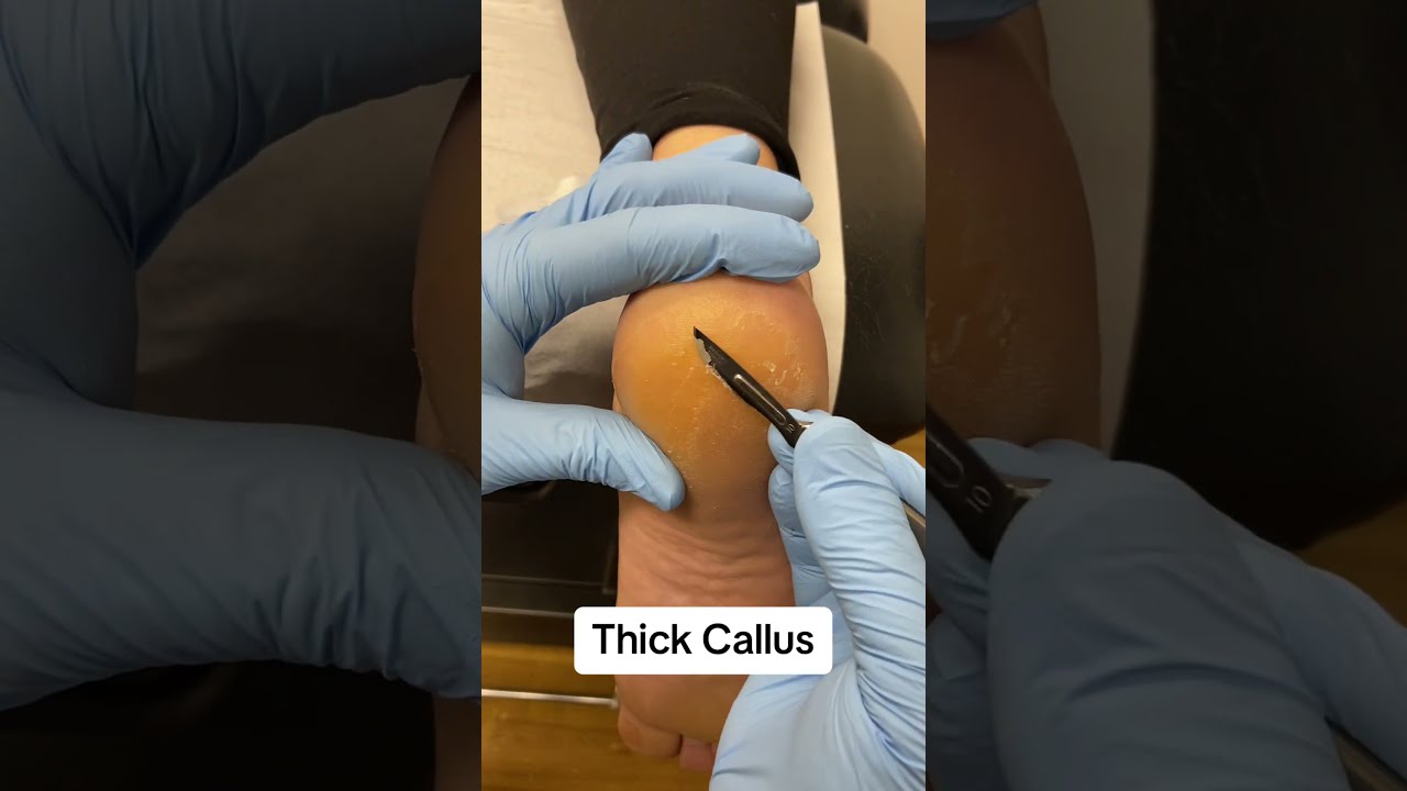
If your ankle Podiatrist Jersey City essexunionpodiatry.com still swells at the end of the day a month after the injury, or if it gives way during daily activities, something is off. Chronic ankle pain often reflects missed diagnoses like peroneal tendon tears, osteochondral lesions, or subtle instability. This is where a comprehensive evaluation by a foot and ankle doctor pays dividends. Expect a repeat exam, functional tests, gait assessment, and likely an MRI if previous imaging has been limited to X-rays.
Some patients carry older foot mechanics that set them up to sprain repeatedly. A high arch foot places more weight laterally, inviting inversion. Flat feet can strain the posterior tibial tendon, shifting load medially and changing how the ankle stabilizes. An orthotic evaluation looks at these patterns. A custom orthotics podiatrist can offload risky areas and steady the foot under the ankle joint. Small changes in alignment produce big differences in confidence on uneven ground.
Special considerations: athletes, kids, and those with medical conditions
For athletes, the clock matters. A running injury podiatrist balances the need to protect tissue with the reality of seasons and competition. A targeted MRI can shorten guesswork when return-to-play decisions loom. That said, most teams prioritize function over images. If you can hop, cut, and land without pain or instability, and your exam is stable, you are more likely ready than a borderline MRI suggests.
In children, growth plate injuries can mimic ligament sprains, and tenderness over the growth plate should prompt X-rays even if weight bearing is tolerable. Kids heal quickly, but they also need guardrails to avoid reinjury. A children’s foot doctor will also look for hypermobility syndromes that make recurrent sprains more likely, and offer strengthening and bracing strategies that match active lives.
For people with diabetes, neuropathy, or vascular disease, the threshold for imaging is lower. An ankle sprain that seems routine can mask a fracture or evolve into Charcot changes in those with severe neuropathy. A foot circulation doctor or wound care podiatrist considers skin integrity and swelling in brace selection. If a boot rubs and a callus forms, an ulcer can follow. Early check-ins prevent spirals that are far harder to reverse.
Costs, insurance, and smart sequencing
Many patients ask about cost. X-rays are the least expensive and usually approved without fuss. MRI costs vary widely by region and facility. Hospital-based MRIs often list higher than outpatient centers. If insurance requires prior authorization, having a clear exam note that documents failed conservative care and specific concerns speeds approval. As a podiatry care provider, I explain why we are ordering a test and what we hope to learn. If an MRI would not change the plan, I say so and offer an alternative path.
There is no shame in a phased approach: start with X-rays, treat the likely sprain, reassess at the two-week mark, then escalate to MRI if recovery stalls or the exam localizes to structures that plain films cannot show. That path uses resources wisely while still respecting the possibility of hidden problems.
What recovery looks like when imaging guides the path
Ankle sprains that stay inside the mild to moderate range often return to full activity within 2 to 6 weeks with proper rehab. High ankle sprains stretch longer, often 6 to 12 weeks, because the syndesmosis stabilizes rotation rather than simple side-to-side motion. Tendon tears may require immobilization for 4 to 6 weeks, then a graded strengthening program that keeps symptoms at bay while load increases. Osteochondral lesions vary: small, stable lesions may heal with offloading and time, while larger, unstable ones may warrant a foot and ankle surgeon’s input.
Imaging sharpens those timelines. A clear X-ray with a classic low ankle sprain lets us push rehab confidently. An MRI that shows a bone bruise under the talar dome tells us to respect impact for a few extra weeks, even if the ankle “feels okay” on flat ground. That nuance prevents setbacks.
When surgery enters the conversation
Surgery is not the first move for sprains, but it has a firm place for specific problems. Chronic ankle instability that fails a well-run rehabilitation program can benefit from ligament reconstruction, often performed by a podiatric foot surgeon or a foot and ankle surgeon with a focus on ligament procedures. Displaced fractures, unstable syndesmotic injuries, and large, unstable osteochondral lesions are better served by surgical stabilization. MRI and CT together outline the roadmap for these cases.
Minimally invasive foot surgeons increasingly use smaller incisions and arthroscopy to address cartilage lesions, debride scar tissue, or evaluate the joint directly. Not every case fits a minimally invasive approach, but when it does, recovery can be smoother with less soft tissue disruption. As always, the decision weighs anatomy, activity goals, and the patient’s tolerance for risk and downtime.
How to think about your next step if you are unsure
If you are standing at the crossroads and are unsure whether to push for an MRI or to stay the course, ask two questions. First, what specific diagnosis are we considering, and would an MRI confirm or exclude it? Second, if the MRI is positive for that diagnosis, would treatment change today? If both answers are clear, the test is often justified. If the answers are fuzzy, a reassessment in one to two weeks with a focused exam may be the smarter path.
The right clinician helps you navigate these choices. A podiatry specialist who sees ankle injuries daily will make imaging decisions feel less like guesswork and more like a guided process. That is the real value of seeing an ankle injury specialist rather than treating every sprain the same.
Final takeaways you can act on now
- Get an X-ray right away if you cannot take four steps, have bone tenderness along the back edge of the malleoli, or have significant pain at the base of the fifth metatarsal. Consider MRI if pain and function are not improving after 10 to 14 days, or sooner if a high ankle sprain, tendon tear, or osteochondral lesion is suspected. Use imaging to answer a decision, not to collect trivia. If the result would not change treatment, timing the test later is reasonable. Rehab early, but protect the ankle from motions that reproduce sharp pain. Progress balance and strength in a graded fashion. If your story includes diabetes, neuropathy, recurrent sprains, or elite sport demands, involve a foot and ankle specialist early. The threshold for imaging is different.
Ankles recover best when care matches the injury’s true nature. A medical foot doctor who listens, examines carefully, and orders the right test at the right time can save you months of frustration. Whether you need the straightforward reassurance of a normal X-ray or the deeper view an MRI offers, the goal is the same: stable footing, confident movement, and a return to what you love without looking down at every step.